Molt stage impacts mating in closed-thelycum species, market value, ablation
The molting cycle influences many aspects of crustacean biology, including animal morphology, cellular metabolism, physiology, and behavior. In shrimp culture, many production aspects are also related to molting, such as mating in closed-thelycum species, market value (affected by exoskeleton hardness), and successful eyestalk ablation to induce ovarian development in broodstock animals. In addition, because of the high prices paid for live shrimp in some markets, it is important to plan the harvest operation to achieve the best survival rate.
The ability to accurately determine the molting stage in cultured shrimp populations can be a highly useful management tool.
Crustacean growth and molting
Growth in crustaceans is not a continuous process. Decapod crustaceans must first loosen the connections between their living tissue and the cuticle. Then they must move out relatively rapidly from the confines of this cuticle, take up water to expand the new, flexible exoskeleton, and quickly harden it so it provides protection and support for locomotion.
The actual act of shedding the old exoskeleton, ecdysis, is the most obvious manifestation of the molt cycle. However, it comprises only a few minutes of a cycle that in some crustacean species takes a year or more to complete, and is divided into several major stages with numerous substages.
Penaeid molting stages
Penaeid shrimp molt at intervals of a few days or weeks. Their molt cycle is divided into six stages: early postmolt (stage A), late postmolt (stage B), intermolt (stage C), early premolt (stage D0-D1), late premolt (stage D2-D3), and ecdysis (stage E). Available literature on the molt staging of penaeid shrimp is somewhat difficult to interpret and generally not useful for practical applications.
In 1987, Robertson et al. described a straightforward method to determine the molt stages in the Journal of the World Aquaculture Society. The method was based on the morphological changes in the uropods. However, clear images of these morphological changes were not available.
A recent study by the authors was designed to produce clear illustrations of the major molt stages and ecdysis that could be used as a reference to readily determine molt stage.
Shrimp acclimation
Live 30- to 50-gram cultured Kuruma prawns, (Marsupenaeus japonicus), from Acquatina Lake in Frigole, Lecce, Italy, (location of the Marine Aquaculture and Fisheries Research Centre) were acclimated in three sand-bottom aquariums under controlled conditions for one week before the experiment. The general health of the animals was assessed daily, and any dead animals and shrimp shells were recorded and removed along with leftover food. Water chemical and physical parameters were maintained within optimal ranges, and no evidence of disease was observed.
Observation results
To perform the molt staging, shrimp uropods were placed gently on a glass slide and observed at 10x magnification under an optical microscope equipped with a photo camera. Pictures were taken of the internal uropod morphology at each of the molting stages.
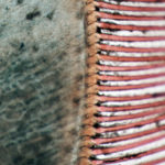
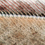
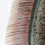
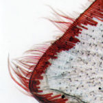
In stage A (Fig. 1), which occurred immediately after ecdysis, a pigmented cellular matrix completely filled the setal bases. During stage B (Fig. 2), the cellular matrix retracted from the setal base and a clear space was easily recognized in the bases. In stage C (Fig. 3), the matrix was absent from the setal bases and the pigment seemed to form an epidermal line at the bases of the setal nodes.
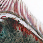
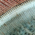
At the D0-D1 stage (Fig. 4), the pigment retracted from the bases of the setal nodes, leaving the old cuticle. During D2-D3 (Fig. 5), the new developing setae were observed. Stage E, the actual shedding of the exoskeleton, occurred at night in less than one minute, which made it very difficult to find animals in this molting stage. Fig. 6 shows the new setae that were extruded from the matrix.
Conclusion
For various activities in shrimp culture operations, it is important to correctly determine the molting stages of animals. The high-quality illustrations produced during this study provide a practical visual aid that can be used to readily determine the molt stages of shrimp.
(Editor’s Note: This article was originally published in the October 2003 print edition of the Global Aquaculture Advocate.)
Now that you've reached the end of the article ...
… please consider supporting GSA’s mission to advance responsible seafood practices through education, advocacy and third-party assurances. The Advocate aims to document the evolution of responsible seafood practices and share the expansive knowledge of our vast network of contributors.
By becoming a Global Seafood Alliance member, you’re ensuring that all of the pre-competitive work we do through member benefits, resources and events can continue. Individual membership costs just $50 a year.
Not a GSA member? Join us.
Authors
-
Dr. Loredana Zilli
Laboratory of Comparative Physiology
Department of Biological and Environmental Sciences and Technologies
University of Lecce
Via Provinciale Lecce-Monteroni 73100 Lecce, Italy -
Dr. Roberta Schiavone
Laboratory of Comparative Physiology
Department of Biological and Environmental Sciences and Technologies
University of Lecce
Via Provinciale Lecce-Monteroni 73100 Lecce, Italy -
Prof. Sebastiano Vilella
Laboratory of Comparative Physiology
Department of Biological and Environmental Sciences and Technologies
University of Lecce
Via Provinciale Lecce-Monteroni 73100 Lecce, Italy -
Dr. Giuseppe Scordella
Marine Aquaculture and Fisheries Research Centre
Frigole-Lecce, Lecce, Italy -
Dr. Vincenzo Zonno
Marine Aquaculture and Fisheries Research Centre
Frigole-Lecce, Lecce, Italy
Tagged With
Related Posts
Health & Welfare
A holistic management approach to EMS
Early Mortality Syndrome has devastated farmed shrimp in Asia and Latin America. With better understanding of the pathogen and the development and improvement of novel strategies, shrimp farmers are now able to better manage the disease.
Intelligence
A land grab for salmon (and shrimp) in upstate New York
The operators of Hudson Valley Fish Farm see their inland locale as a pilot to prove that land-based fish farming, located in close proximity to major metropolitan markets, can be successful.
Health & Welfare
A study of Zoea-2 Syndrome in hatcheries in India, part 1
Indian shrimp hatcheries have experienced larval mortality in the zoea-2 stage, with molt deterioration and resulting in heavy mortality. Authors investigated the problem holistically.
Health & Welfare
Stirling researcher preps eyestalk-ablation alternative trials
University of Stirling Ph.D. student Simão Zacarias, who is from is Beira, Mozambique, will soon travel to Isla del Tigre, Honduras, to document evidence showing the benefits of breeding shrimp without eyestalk ablation. His is a journey of hopeful discovery.


